|
READING
The Eye’s Optical Mechanism
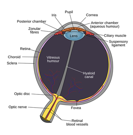
At the start of their journey through the eye’s optical system, electromagnetic rays encounter the cornea, which is a transparent, convex, aspheric membrane covering the iris and pupil.
VIDEO: A Journey through the Eye (2:38)
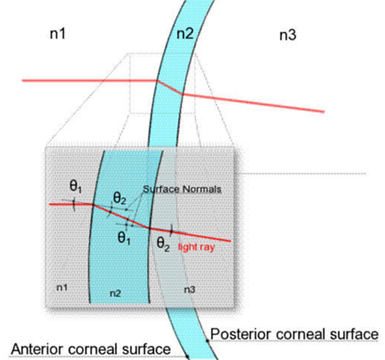
The primary task of the cornea is to direct incoming rays towards the pupil. This is accomplished through refraction. As rays pass through the curved outer and inner surfaces of the cornea, they are refracted, or bent, towards the pupil. The cornea is the most refractive element in the eye, with a refractive power of about 70% of the power of the whole eye.
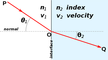
The refractive index of the cornea ranges from 1.373 (corneal stroma, posterior), 1.380 (corneal stroma, anterior) to 1.401 (corneal epithelium). The refractive index of the aqueous humor (the liquid beneath the cornea in front of the pupil) is almost the same: 1.336. The refractive index is greatest, and the most bending occurs, when rays pass from the air through the convex anterior surface of the cornea. The rays are refracted again, but much less so, as they pass from the concave posterior surface of the cornea into the aqueous humor.
Rays entering perpendicular to the central portion of the corneal dome are minimally refracted and pass directly through the centre of the pupil.
After entering the pupil, the rays then pass through the crystalline lens whose curvature, and therefore angle of refraction, is coordinated with the size of the pupil.
The refractive index of the lens varies from 1.3636 (cortex) to 1.4034 (nucleus). The refractive index of the vitreous humor filling the inside of the eyeball is 1.336.
Besides serving to filter out harmful ultraviolet radiation, the primary task of the lens is to distribute and focus incoming rays around the inside surface of the eyeball where the retina is located.
Rays passing through the centre of the lens are refracted to converge precisely on the fovea. All the other incoming rays are distributed over the surrounding surface of the retina. Whereas the image on the fovea is clear and sharp, the image appearing on the rest of the retina is not sharply focused or clearly defined.
The optical system of the eye is often likened to that of a photographic camera. Both make use of an adjustable aperture to control the amount of entering rays and employ a lens to focus these rays onto a light-sensitive surface. A simple comparison, however, shows that the eye is much more complex than a camera.
VIDEO: Eye Works 1 (13:57)
VIDEO: Dua’s Layer (00:59), a newly discovered layer of the cornea
The image on the retina is formed in response to rays entering the eye from a very large field of view. Because the cornea is dome-shaped and itself located upon the curved surface of the eyeball, the horizontal field of view for a forward-pointing human eye is approximately 210 degrees.
For binocular humans, the overlap zone of the horizontal field of view of both eyes is approximately 120 degrees.
The vertical field of view is about 120 degrees. The view is blocked a little on one side by the nose and above by the brow. A standard 50mm lens on a 35mm camera, in contrast, has a horizontal angle of view of only 40 degrees and a vertical angle of view of only 27 degrees.
A standard lens is defined as a lens that has a focal length equal in distance to the diagonal of the negative. It has no telephoto or wide-angle properties and a depth of focus of 10 or 12 feet.
In order to increase (or decrease) the angle of view, different lenses must be used. Using an 18mm ‘wide-angle’ lens, a conventional 35mm camera can achieve a maximum horizontal field of view of 100 degrees, which is still only approximately half that seen by the eye.
A camera could encompass the eye’s field of view only by using a ‘fish-eye’ lens, which can provide an angle of view of 180 degrees or more.
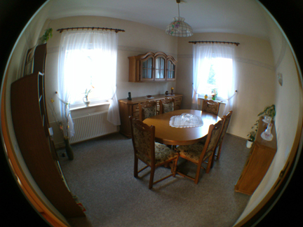
Except for a small area at the centre, however, the image produced by a fish-eye lens is subject to linear distortion (where straight lines are curved) and proportion distortion (where the proportions of objects are exaggerated), especially towards the periphery.
Do similar distortions occur in the eye?
If they do, we may automatically adjust the image to compensate for them, in the same way we somehow adjust for the fact that the image appearing on the retina is upside down and reversed and that part of it is hidden by our blind spot.
It is also possible that the combination of curved surfaces in the eye — from the cornea to the lens to the retina — serves to somehow ‘foreshorten’ the image in such a way that the distortions are optically corrected.
The lens in a camera focuses the entire incoming body of rays onto the photographic film so that the whole image is in focus (within the plane of the field of focus; see below).
In contrast, only a small central portion of what the eye sees is in sharp focus at any given moment. Moreover, whereas the camera lens focuses rays onto a flat surface (the photographic film), the crystalline lens in the eye distributes rays over a concave surface (the retina).
However, it is through these differences that the camera can provide some important clues about human vision.
A key feature of the eye is the way it adjusts for focal attention. As focal attention shifts, changes occur in pupil size and lens shape.
These changes are designed to alter the eye’s depth of focus.
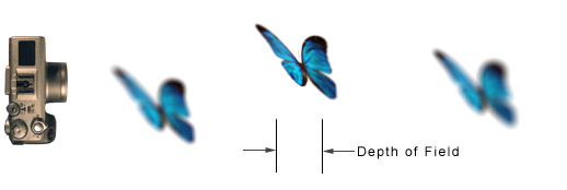
Depth of focus refers to a zone located within the space between the eye and infinity. It is the distance from the nearest to the farthest point within the field of vision where objects are in focus.
Depth of focus can be made to vary from narrow to large. In a camera, depth of focus (or depth of field, which is the term generally preferred by photographers) varies with the size of the lens opening, its focal length, and the distance of the subject focused on.
Focal length is the distance from the optical centre of the lens to the film behind it. The focal length of a lens is commonly given in millimeters (mm).
The depth of field is narrow (or short) if the size of the lens opening is big and the focal length long. Conversely, with the subject distance unchanged, depth of field is made larger (or longer) by decreasing the size of the lens opening and decreasing its focal length.
The eye’s depth of focus is controlled by the size of the pupil and by the shape of the lens. In its ‘normal’ state, the eye has a large depth of focus; the size of the pupil is small and the focal length is short.
Focal length in the eye is the distance from the optical centre of the lens to the fovea located in the retina. The advantage of a large depth of focus is that it places more of the field of view in focus.
The disadvantage is a reduction in the quality of the image. This is due to the smaller pupil size, which reduces the amount of light and therefore the amount of visual information entering the eye.
The reduction in image quality, however, is only slight, and objects within the depth-of-focus zone will look acceptably sharp to the eye.
There are times, though, when something catches your visual attention or stimulates your interest and you want to see it as clearly and as precisely as possible.
As we noted earlier, when interest is stimulated, the size of the pupil enlarges, and at the same time the lens becomes more elongated and flatter.
As the lens elongates, its optical centre is shifted forward, thereby increasing the eye’s focal length. Simultaneously, as the pupil enlarges, more light enters the eye, which results in an increase in image quality.
According to M. Millodot, in The Senses, edited by H. B. Barlow and J. D. Mollon, p. 57, when the pupil diameter is 2 mm, the depth of field at infinity extends to approximately 2.3 m in front of the eye, and at 1 m (for the same pupil size) it varies from approximately 1.8 to 0.7 m. When the size of pupil is 4 mm, the depth of field at infinity extends to about 3.5 m, and at 1 m it varies from approximately 1.4 to 0.8 m.
Image quality is also enhanced because the altered shape of the lens reduces distortion due to spherical aberration.
The advantage of a narrow depth of focus is that it permits a sharper and more focused visual scrutiny of whatever has caught our attention.
The disadvantage is that overall visual awareness is reduced.
This might be dangerous under certain circumstances. Imagine standing in a room crowded with people. As your eyes disinterestedly scan the crowd, a large depth of field permits you to recognize the faces of people both nearby and a little further away.
Suddenly, you see an attractive stranger. Your pupils enlarge, and the eye immediately adjusts to a more narrow depth of focus in order to scrutinize this person more clearly. People standing in front of and behind this person become less distinct. While your attention is focused on this stranger, however, you may fail to notice this person’s companion standing close by staring at you with increasing annoyance!
Our ability to manipulate depth of focus, however, is limited in range. This range is determined mostly by the extent to which the lens can change shape.
When viewing an object close at hand, there is a point where the zonules reach maximum tension and the lens is at its flattest. If the object is brought closer to the eye, depth of focus is lost because the lens is unable to adjust any further.
Conversely, when viewing objects at a distance, depth of focus ceases to function once the zonules have reached minimum tension and the lens is at its most spherical; this occurs at a distance of more than 6 meters (20 feet) away. At distances greater than 6 meters, depth of focus expands to “infinity”. When the eye is focused on “infinity”, depth of focus starts at a point beyond 6 meters and extends to infinity.
The large depth of field also means that the quality of the image is reduced. As the object’s distance from the eye is increased, further deterioration in image quality occurs due to the intervening air or atmosphere, which causes the outlines of the object to become less distinct, details to become less sharp or not discerned at all, and colour saturation and range to be reduced.
Because the lens is at its most spherical, there is no possibility of adjustment in order to focus visual attention on an object lying somewhere within this range. At the same time, when the eye is focused on “infinity”, objects closer than 6 meters fall out of focus.
Indeed, small intervening objects virtually disappear. This was no doubt an advantage to someone hiding in tall grass from a lion in a distant clearing! With the eye focused on the lion at infinity, the intervening grass stalks in front of your face become almost invisible, which permits a fuller view of the lion.
However, without moving, it is possible to shift focus from the lion to the stalks of grass, and back again. This can be a conscious, deliberate decision. In other words, we have some measure of control over what we want to focus on. Focus is not completely “automatic”, a response triggered by blurredness of the perceived object.
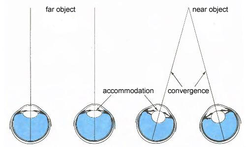
The mechanism controlling accommodation (the changes in the shape of the lens), and thereby depth of field and focus, would appear to be the angle of convergence.
Convergence
Convergence is when both eyeballs are turned so that the visual axis of each eye converges on the object of visual attention. The nearer the object, the more the eyes must turn to focus on it, and therefore the greater the angle of convergence.
The opposite occurs for objects further away. The eyeball is physically rotated or turned in its socket through the action of six muscles attached to its exterior surface.
Convergence and Accommodation
These six extraocular muscles are controlled by the parasympathetic nervous system, that also controls the ciliary muscle (which adjusts the shape of the lens) and the pupillary sphincter muscle (which causes the pupil to become smaller).
All eight muscles are connected to the so-called Edinger-Westphal nucleus located in the midbrain, which coördinates their actions.
Thus, when you look at an object close at hand, motor neurons in the Edinger-Westphal nucleus simultaneously cause the eyes to converge and the lens to accommodate so that the object is brought into focus.
The Edinger-Westphal nucleus also controls the phenomenon of the pupillary light reflex; when a light shines into one eye, the pupils of both eyes will simultaneously contract.
A similar mechanism most likely governs convergence; as one eye catches a point of interest and rotates to look at it, the other eye automatically follows suit.
Convergence also supplies the mechanism for seeing depth and judging distance (called stereopsis). When looking at an object close at hand, the eyeballs turn so that the line of sight converges on the object.
When the object is farther away, the angle of convergence decreases.
From the angle of convergence, the brain can calculate distance, but only in respect of one object relative to another. In order to calculate distances of several objects simultaneously, the brain analyzes disparity.
When viewing a distant object, the image seen in one eye is slightly different from the image seen in the other eye.
You can verify this by alternately closing one eye and then the other. You will notice, as did Leonardo da Vinci when looking at a sphere, that you can see slightly farther around the left side of the sphere with the left eye and slightly farther around the right side with the right eye.
Although Leonardo failed to ponder the consequences of this observation, the British physicist Sir Charles Wheatstone (1802-75) was able to use it to demonstrate the principle of stereopsis by which the brain is able to estimate size and depth by comparing the slightly different image (or disparity) received from each eye.
This works, however, only with objects up to 300 feet (100 meters) away. Beyond that, we have no stereoscopic vision.
|

 ARTH117
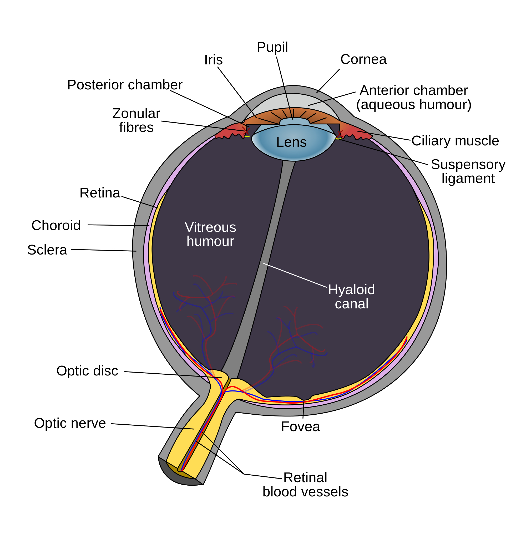
