|
READING
From Eye to Brain
In his Natural History, Pliny (23–79 CE) reports that the “most learned authorities state that the eyes are connected with the brain by a vein”.
Writing in the 2nd century CE, Galen was struck by how the “net-like” retina looked like part of the brain: “if you strip it off entirely and try to gather the whole part together, it will appear to you as brain.”
Galen saw this as evidence of the eye’s superiority over the other sense organs. He declared that, “You will not find the actual substance of the brain in any other sense organ,” and concluded that it showed that the eye “has also a greater share in the nature of the brain.”
Despite Johannes Kepler’s discovery in 1604 that the image was formed on the retina, there was evidently little interest among physiologists in the 17th and 18th centuries in investigating the retina’s connection to the brain.
Early in the 19th century, however, the German physiologist and anatomist Johannes Müller (1801-1858) developed the law of specific nerve energies in which he showed that the sense organ registers only nerve impulses, which are then interpreted by the brain.
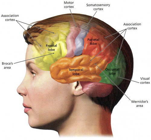
In 1855, the Italian physiologist and anatomist Bartolomeo Panizza (1785-1867) published Osservazioni sul nervo ottico (“Observations on the Optic Nerve”) in which he macroscopically traced the visual projection from the optic nerve to the cerebral structures thereby establishing that the visual function was localized in the visual cortex in the occipital region of the posterior cortex of the brain.
A vast amount of research since then has uncovered, and continues to uncover, in greater detail the paths by which optical information from the eyes is transmitted to the primary visual cortex.
The rods and cones in the retina translate the energy carried by individual photons into chemical messages, which are then conveyed out of the eye along the optic nerve.
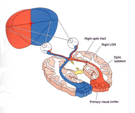
The fibers of the optic nerve from each eye first pass through the optic chiasm (from the Greek letter chi denoting the letter “X”).
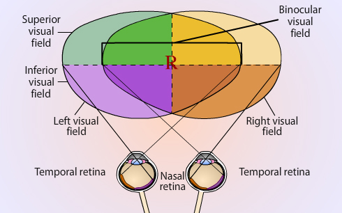
At the optic chiasm, half the fibers from each eye go to one side of the brain and the other half go to the other side. Information is evenly divided between both eyes.
Fibers from the left half of the left retina, and fibers from the left half of the right retina, go to the geniculate body on the left side of the brain.
Fibers from the right half of the left retina, and from the right half of the right retina, go to the right geniculate body.
Most of the one and a half million cells in each lateral geniculate body, located on either side of the brain stem, connect to various areas in the primary visual cortex (or striate cortex) at the rear of the brain.
Some information is also conveyed to and from the superior colliculus, located in the brain stem, which controls eye movements.
These connections from eye to brain are topographically organized, which means that an optic nerve from a particular point in the retina travels to a corresponding point in the lateral geniculate body and thence to a particular point in the primary visual cortex.
What we see with our eyes is thus systematically transmitted from the retina to the geniculate and thence to the cortex.
Because the lens of the eye reverses the image in the retina, light coming from the right in the visual field is projected onto the left side of the retina in each eye and is sent to the left side of the brain; and conversely on the left.
Neurobiological research has shown that visual data is divided into different parts, which are channeled to different areas or “modules” of the visual cortex.
Neurons in the area known as V3, for example, are very sensitive to the orientation of straight lines and edges and the detection of shapes.
Colour and relative lightness and darkness are processed in the area known as V4.

These basic visual elements — lines, edges, shapes, colour, lightness, and darkness — constitute what you see at the outset of vision.
|

 ARTH117