|
READING
Retina
Through dissection of the eyeball, the ancient Greek physician and surgeon Galen (129–c. 200/c. 216 CE) had found two moist spherical bodies, “one as soft as moderately melted glass [hyalos], the other as hard as moderately frozen ice [krystallos]”.
The former is the jelly-like filling of the eyeball (later called the vitreous humour, which is contained within a transparent sac called the hyaloid membrane). The latter is the “crystalline” body (later labeled the crystalline humor, and today called the lens), which he observed, “has the shape of a slightly flattened sphere”.
The crystalline body was attached to a “flattened nerve” that resembled “a casting-net”, which caused it to be called “the net-like tunic” or retina.
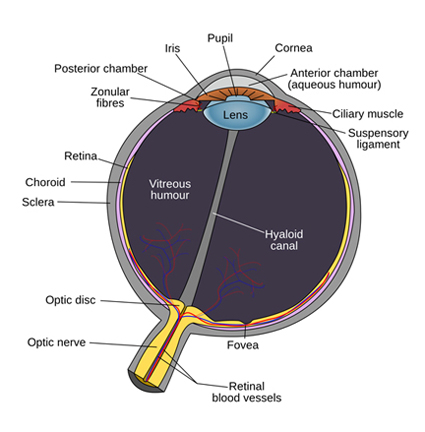
The retina is composed of four microscopically thin layers of cells that line most of the inside of the eyeball. The first layer, next to the choroid, is the epithelium upon which sits a layer composed of some 125 million photon-receptors in the form of long slender rods and short tapered cones.
It is the rods and cones that translate the energy carried by individual photons into chemical messages, which are transmitted to the neurons in two layers of cells that lie above them.
The first layer contains three types of cells: bipolar cells, horizontal cells, and amacrine cells. Above this layer is another containing the retinal ganglion cells.
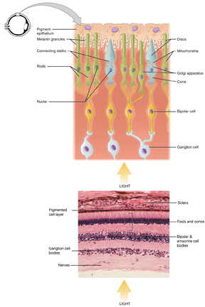
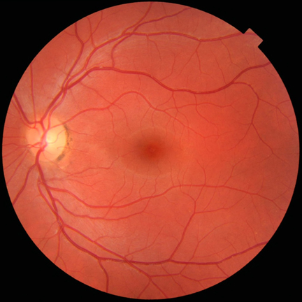
Although both these layers of cells are fairly transparent, they must nevertheless affect the passage of light through them to the rods and cones below. Moreover, above these two layers is a network of veins and arteries, which must also inhibit the passage of light to the rods and cones below.
There seems to be no explanation for this upside-down arrangement. As a result, a large portion of what our eyes see, as we gaze out into the world, is in fact blurred.
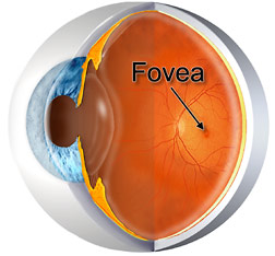
Most of the time we are not really aware of this because we tend to concentrate on looking, which is conducted through a tiny part of the retina called the fovea (at the center of the macula) where vision is at its most acute.
How the Eye Works and the Retina (video)
The fovea is a shallow pit in the retina about one millimeter across located opposite the lens. It occupies an area to the left of the nasal hemifield on the same horizontal meridian that passes through the optic disc, which is where the optic nerve leaves the eye.
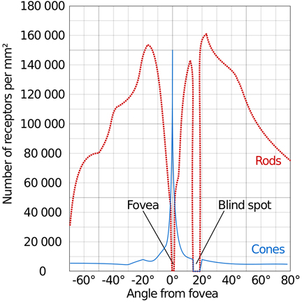
Where the optic nerve exits the eye there are no rods and cones and thus no registration of the light waves hitting this spot on the retina. The result is a blind spot which the brain compensates for by “filling in” the missing information. It does this so effectively that most of the time we are unaware of this small hole in our vision.
The fovea has no cells or blood vessels covering it and is thus directly exposed to incoming electromagnetic radiation.
The axons of the ganglion cells traverse the surface of the retina to gather into a bundle of fibers that leave the eye as the optic nerve.
The connections between rods and cones to bipolar cells and to ganglion cells vary over the surface of the retina.
In or near the fovea, the connection is direct in that a single cone feeds a single bipolar cell, and a single bipolar cell in turn feeds into a single ganglion cell.
Towards the periphery of the retina, however, the connection between cells becomes less direct.
Moving away from the fovea, more rods and more cones converge on more bipolar cells, and more bipolar cells converge on more ganglion cells.
Visual acuity is therefore highest in the area of the fovea and deteriorates as it moves away towards the periphery of the retina.
The cones of the retina have three light-sensitive receptors that are attuned to respond to electromagnetic wavelengths that we sense as the colours red, green, and blue.
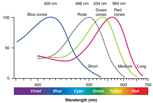
Cones respond only to light at normal intensity and permit us to see colours.
Cones are normally one of the three types, each with different photosins or pigment (the three types make most humans trichromatic.
The response curve is different in each one and thus each responds in different ways to variation in colour.
Rods, on the other hand, are responsible for vision in dim light (“twilight vision”) and provide vision only in shades of gray.
Unlike cones, which have three different types of pigment, all rods have a single light-sensitive pigment.
Rods are far more numerous than cones except in the area of the fovea, which contains only cones. Our eyes automatically resort to rod vision in darkened places (called scotopic vision).
|